Class 12 Chemistry Biomolecules Notes
- Carbohydrates
- Monosaccharides
- Glucose & Fructose
- Disaccharides
- Polysaccharides
- Proteins
- Enzymes
- Vitamins
- Nucleic Acids
- Hormones
- Chapter Summary
- Important Differences
- Exam Questions
- Mind Map
Part 1: Carbohydrates
Chapter Overview
Biomolecules are the chemical substances that make up living organisms. Among them, carbohydrates are the most abundant organic compounds found in nature. They are produced mainly by green plants during photosynthesis and serve as the primary source of energy for almost all living organisms.
1. What are Carbohydrates?
Definition
Carbohydrates are polyhydroxy aldehydes, polyhydroxy ketones, or compounds that produce them on hydrolysis.
They are commonly known as sugars or saccharides.
Important Point
Earlier, carbohydrates were believed to be hydrates of carbon because many have the general formula:
Cx(H₂O)y
However, this formula is not a true definition because:
- Some compounds satisfy the formula but are not carbohydrates.
- Some carbohydrates do not follow this formula.
Examples
| Carbohydrate | Common Name |
|---|---|
| Glucose | Blood sugar |
| Fructose | Fruit sugar |
| Sucrose | Cane sugar |
| Lactose | Milk sugar |
| Maltose | Malt sugar |
| Starch | Plant storage food |
| Cellulose | Plant cell wall material |
| Glycogen | Animal starch |
Why are Carbohydrates Important?
Carbohydrates perform several essential functions.
1. Main Source of Energy
- Glucose is the body’s preferred energy source.
- Cells break down glucose during respiration to produce ATP.
2. Energy Storage
Plants store carbohydrates as:
➡ Starch
Animals store carbohydrates as:
➡ Glycogen
3. Structural Role
Cellulose forms the rigid cell wall of plants.
4. Industrial Importance
Carbohydrates are used in making:
- Paper
- Cotton cloth
- Textiles
- Wood products
- Brewing industries
5. Biological Importance
Certain sugars like ribose and deoxyribose are components of nucleic acids (RNA and DNA).
Classification of Carbohydrates
Carbohydrates are classified according to the products formed during hydrolysis.
                Carbohydrates
                      │
      ┌───────────────┼───────────────┐
      │               │               │
Monosaccharides   Oligosaccharides  Polysaccharides
1. Monosaccharides
Definition
Monosaccharides are the simplest carbohydrates that cannot be hydrolysed into smaller carbohydrate units.
These are also called simple sugars.
Characteristics
- Sweet in taste
- Water soluble
- Colourless crystalline solids
- Reducing sugars
- Immediate source of energy
Examples
- Glucose
- Fructose
- Ribose
- Galactose
Classification of Monosaccharides
Based on Functional Group
| Type | Functional Group | Example |
|---|---|---|
| Aldose | Aldehyde (-CHO) | Glucose |
| Ketose | Ketone (>C=O) | Fructose |
Based on Number of Carbon Atoms
| Carbon Atoms | Name |
|---|---|
| 3 | Triose |
| 4 | Tetrose |
| 5 | Pentose |
| 6 | Hexose |
| 7 | Heptose |
Examples
- Ribose → Pentose
- Glucose → Hexose
- Fructose → Ketohexose
2. Oligosaccharides
Definition
Carbohydrates that produce 2–10 monosaccharide molecules on hydrolysis are called oligosaccharides.
The most common members are disaccharides.
Examples
- Sucrose
- Maltose
- Lactose
3. Polysaccharides
Definition
Polysaccharides produce a large number of monosaccharides when hydrolysed.
Characteristics
- Not sweet
- Generally insoluble in water
- High molecular weight
- Storage and structural carbohydrates
Examples
- Starch
- Cellulose
- Glycogen
Classification Based on Reducing Property
Carbohydrates are also classified as reducing and non-reducing sugars.
Reducing Sugars
These reduce:
- Fehling’s solution
- Tollens’ reagent
Examples:
- Glucose
- Fructose
- Maltose
- Lactose
Non-Reducing Sugars
Do not reduce these reagents.
Example:
- Sucrose
Quick Comparison Table
| Property | Monosaccharides | Oligosaccharides | Polysaccharides |
|---|---|---|---|
| Hydrolysis | Cannot be hydrolysed | Gives 2–10 units | Gives many units |
| Sweet Taste | Yes | Usually yes | No |
| Solubility | High | High | Low |
| Molecular Size | Small | Medium | Very large |
| Examples | Glucose | Sucrose | Starch |
NCERT Important Points
✔ Carbohydrates are mainly produced by plants.
✔ Chemically, carbohydrates are polyhydroxy aldehydes or ketones.
✔ Monosaccharides are reducing sugars.
✔ Polysaccharides are also called non-sugars because they are not sweet.
✔ Starch is the storage carbohydrate of plants.
✔ Glycogen is the storage carbohydrate of animals.
✔ Cellulose is the main component of plant cell walls.
Memory Tricks
Types of Carbohydrates
MOP
- M → Monosaccharides
- O → Oligosaccharides
- P → Polysaccharides
Storage Forms
Plant → Starch
Animal → Glycogen
Structural Carbohydrate
Cellulose → Cell Wall
Part 2: Monosaccharides — Glucose and Fructose
1. Glucose
Introduction
Glucose is one of the most important monosaccharides.
Common Names:
- Blood sugar
- Dextrose
Classification:
Glucose is:
- A monosaccharide
- An aldohexose
- A reducing sugar
- A member of the D-series
Molecular Formula:
C6H12O6
Glucose occurs naturally in:
- Sweet fruits
- Honey
- Ripe grapes
It is also a basic unit of many carbohydrates such as:
- Starch
- Cellulose
2. Preparation of Glucose
(A) From Sucrose (Cane Sugar)
When sucrose is heated with dilute mineral acids like:
- HCl
- H₂SO₄
it undergoes hydrolysis and produces equal amounts of:
- Glucose
- Fructose
Reaction:
Sucrose + Water → Glucose + Fructose
(B) From Starch
Commercial glucose is prepared by hydrolysis of starch.
Conditions:
- Dilute H₂SO₄
- Temperature: 393 K
- Pressure: 2–3 atm
Reaction:
Starch + Water → Glucose
3. Structure of Glucose
The structure of glucose was determined by studying its chemical reactions.
Evidence for Open Chain Structure
1. Molecular Formula
The molecular formula of glucose was found to be:C6H12O6
This indicates the presence of six carbon atoms.
2. Formation of n-Hexane
On prolonged heating with HI:
Glucose gives n-hexane.
Conclusion:
All six carbon atoms are arranged in a straight chain.
3. Presence of Carbonyl Group
Glucose reacts with:
- Hydroxylamine
- Hydrogen cyanide (HCN)
to form addition products.
Conclusion:
Glucose contains a carbonyl group (>C=O).
4. Presence of Aldehyde Group
Glucose reacts with bromine water and gets oxidised to:
Gluconic acid
Conclusion:
The carbonyl group in glucose is an aldehyde group.
Therefore glucose is an aldohexose.
5. Presence of Five Hydroxyl Groups
Glucose reacts with acetic anhydride to form:
Glucose pentaacetate
Conclusion:
Glucose contains five –OH groups.
6. Presence of Primary Alcohol Group
Oxidation of glucose with nitric acid gives:
Saccharic acid
Conclusion:
Glucose contains a primary alcoholic group.
Open Chain Structure of Glucose
According to Fischer projection:
        CHO
         |
    H — C — OH
         |
    OH— C — H
         |
    H — C — OH
         |
    H — C — OH
         |
       CH2OH
This structure explains many properties of glucose.
However, some observations could not be explained by this structure.
4. Limitations of Open Chain Structure
The open-chain structure failed to explain:
(1) Absence of Aldehyde Reactions
Although glucose contains an aldehyde group, it:
- Does not give Schiff’s test
- Does not form sodium bisulphite addition product
(2) No Reaction of Pentaacetate with Hydroxylamine
Glucose pentaacetate does not react with hydroxylamine.
This suggests that a free aldehyde group is absent.
(3) Existence of Two Forms
Glucose exists in two crystalline forms:
α-glucose
- Melting point: 419 K
β-glucose
- Melting point: 423 K
The open-chain structure cannot explain these two forms.
5. Cyclic Structure of Glucose
To explain these observations, it was proposed that glucose forms a cyclic structure.
Ring Formation
The –OH group at carbon number 5 attacks the aldehyde group at carbon number 1.
This produces a cyclic hemiacetal.
Important:
Glucose forms a six-membered ring.
This structure is called:
Pyranose Structure
because it resembles the cyclic compound pyran.
6. Anomers of Glucose
The two cyclic forms of glucose differ only in the arrangement of the –OH group at carbon-1.
Carbon-1 is called:
Anomeric Carbon
The two forms are:
α-D-Glucose
- OH group at C-1 has one orientation
β-D-Glucose
- OH group at C-1 has opposite orientation
These two forms are called:
Anomers
Definition:
Isomers that differ only in the configuration around the anomeric carbon are called anomers.
7. D and L Configuration of Glucose
The letters D and L represent the configuration of a molecule.
They are not related to:
- (+) or (–) optical rotation
D-Configuration:
The –OH group on the lowest asymmetric carbon is on the right side.
L-Configuration:
The –OH group is on the left side.
Glucose has:
D-(+)-Glucose
Meaning:
- D → Configuration
- (+) → Dextrorotatory nature
8. Fructose
Introduction
Fructose is another important monosaccharide.
Common Name:
Fruit sugar
Sources:
- Fruits
- Honey
- Vegetables
Classification:
Fructose is:
- A monosaccharide
- A ketohexose
- A reducing sugar
- A member of D-series
Molecular Formula:
C6H12O6
Fructose is written as:
D-(–)-Fructose
because it is laevorotatory.
9. Structure of Fructose
Fructose contains:
- Six carbon atoms
- Ketone group at carbon number 2
Therefore, it is a:
Ketohexose
Cyclic Structure of Fructose
Fructose forms a cyclic structure by reaction between:
- C-5 hydroxyl group
- C-2 keto group
This forms a:
Five-membered ring
called:
Furanose Structure
because it resembles furan.
Glucose vs Fructose (Important Difference)
| Property | Glucose | Fructose |
|---|---|---|
| Formula | C₆H₁₂O₆ | C₆H₁₂O₆ |
| Type | Aldohexose | Ketohexose |
| Functional group | Aldehyde | Ketone |
| Carbonyl position | C-1 | C-2 |
| Common name | Dextrose | Fruit sugar |
| Optical nature | D-(+) | D-(–) |
| Ring structure | Pyranose | Furanose |
Important Exam Points ⭐
Remember:
Glucose
- Aldohexose
- Pyranose ring
- Six-membered cyclic structure
- Forms α and β anomers
Fructose
- Ketohexose
- Furanose ring
- Five-membered cyclic structure
Part 3: Disaccharides and Polysaccharides
1. Disaccharides
Definition
Disaccharides are carbohydrates that produce two monosaccharide molecules on hydrolysis.
They are formed when two monosaccharide units join together by a bond called:
Glycosidic Linkage
Glycosidic Linkage
Definition:
A glycosidic linkage is an oxygen bridge formed between two monosaccharide molecules by the loss of a water molecule.
Formation:
Monosaccharide + Monosaccharide → Disaccharide + Water
Example:
Glucose + Fructose → Sucrose + H₂O
Reducing and Non-Reducing Disaccharides
Disaccharides are classified according to the presence of free aldehyde or ketone groups.
1. Reducing Sugars
Definition:
Disaccharides that contain a free reducing group are called reducing sugars.
They can reduce:
- Fehling’s solution
- Tollens’ reagent
Examples:
- Maltose
- Lactose
2. Non-Reducing Sugars
Definition:
Disaccharides in which reducing groups are involved in glycosidic bond formation are called non-reducing sugars.
Example:
- Sucrose
2. Sucrose
Introduction
Sucrose is one of the most common disaccharides.
Common Name:
Cane sugar
Sources:
- Sugar cane
- Sugar beet
Composition
Sucrose is made up of:
- One molecule of α-D-glucose
- One molecule of β-D-fructose
Hydrolysis:
Sucrose + Water → Glucose + Fructose
Glycosidic Bond in Sucrose
The linkage occurs between:
- C-1 of α-D-glucose
- C-2 of β-D-fructose
Because both reducing groups participate in bond formation:
➡ Sucrose is a non-reducing sugar.
Invert Sugar
Sucrose is:
- Dextrorotatory (+)
After hydrolysis:
- Glucose → Dextrorotatory (+)
- Fructose → Laevorotatory (–)
Since fructose has stronger negative rotation, the mixture becomes:
➡ Laevorotatory
This hydrolysed product is called:
Invert Sugar
3. Maltose
Introduction
Maltose is a disaccharide consisting of:
Two α-D-glucose units
Linkage
The two glucose molecules are joined by:
α(1 → 4) glycosidic linkage
Meaning:
- C-1 of first glucose joins with
- C-4 of second glucose
Reducing Nature
The second glucose molecule has a free aldehyde group.
Therefore:
➡ Maltose is a reducing sugar.
4. Lactose
Introduction
Lactose is commonly known as:
Milk Sugar
It is found in milk.
Composition
Lactose contains:
- β-D-galactose
- β-D-glucose
Linkage
The bond is between:
- C-1 of galactose
- C-4 of glucose
Reducing Nature
A free aldehyde group can develop in glucose.
Therefore:
➡ Lactose is a reducing sugar.
Comparison of Disaccharides
| Property | Sucrose | Maltose | Lactose |
|---|---|---|---|
| Common name | Cane sugar | Malt sugar | Milk sugar |
| Components | Glucose + Fructose | Glucose + Glucose | Galactose + Glucose |
| Linkage | α-D-glucose C1 → β-D-fructose C2 | α-glucose C1 → glucose C4 | β-galactose C1 → glucose C4 |
| Reducing nature | Non-reducing | Reducing | Reducing |
| Source | Sugar cane | Barley/malt | Milk |
5. Polysaccharides
Definition
Polysaccharides are carbohydrates that produce a large number of monosaccharide units on hydrolysis.
They are polymers of monosaccharides joined through glycosidic linkages.
Characteristics
- High molecular mass
- Generally tasteless
- Insoluble or slightly soluble in water
- Used mainly for storage and structural purposes
Examples:
- Starch
- Cellulose
- Glycogen
6. Starch
Introduction
Starch is the main storage polysaccharide of plants.
Sources:
- Cereals
- Roots
- Tubers
- Vegetables
It is the most important carbohydrate in the human diet.
Composition of Starch
Starch is made up of:
- α-D-glucose units
It has two components:
(A) Amylose
Properties:
- Water soluble
- About 15–20% of starch
- Long unbranched chain
Linkage:
α-D-glucose units joined by:
C-1 → C-4 glycosidic linkage
(B) Amylopectin
Properties:
- Insoluble in water
- About 80–85% of starch
- Branched structure
Linkages:
Main chain:
C-1 → C-4 linkage
Branching:
C-1 → C-6 linkage
7. Cellulose
Introduction
Cellulose is:
- The most abundant organic substance in plants
- A major component of plant cell walls
Structure
Cellulose is made of:
β-D-glucose units
joined by:
β(1 → 4) glycosidic linkage
Important Point
Humans cannot digest cellulose because our digestive system lacks the enzyme required to break β-glycosidic bonds.
8. Glycogen
Introduction
Glycogen is the storage carbohydrate of animals.
It is also called:
Animal Starch
Location in Body
Glycogen is stored mainly in:
- Liver
- Muscles
- Brain
Structure
Glycogen resembles amylopectin but:
- Has more branches
- Breaks down to release glucose when required
Starch vs Cellulose vs Glycogen
| Feature | Starch | Cellulose | Glycogen |
|---|---|---|---|
| Found in | Plants | Plants | Animals |
| Function | Energy storage | Structural support | Energy storage |
| Monomer | α-glucose | β-glucose | α-glucose |
| Linkage | α(1→4), α(1→6) | β(1→4) | α(1→4), α(1→6) |
| Branching | Amylopectin branched | No branching | Highly branched |
Part 4: Proteins — Amino Acids, Peptide Bonds and Protein Structure
1. Proteins
Introduction
Proteins are among the most important biomolecules present in living organisms.
The word protein comes from the Greek word:
“Proteios”
meaning primary or of prime importance.
Proteins are essential for:
- Growth of the body
- Repair of tissues
- Maintaining body functions
- Structural activities
- Biological reactions
Sources of Proteins
Important sources include:
- Milk
- Cheese
- Pulses
- Peanuts
- Fish
- Meat
Basic Structure of Proteins
Proteins are polymers of α-amino acids.
This means:
- Amino acids are the building blocks.
- Many amino acids join together to form proteins.
2. Amino Acids
Definition
Amino acids are organic compounds containing:
- Amino group (–NH₂)
- Carboxyl group (–COOH)
General structure:
        H
        |
H₂N — C — COOH
        |
        R
Here:
R = side chain
The nature of the R group determines the properties of the amino acid.
Classification of Amino Acids
Amino acids are classified based on:
- Number of amino and carboxyl groups
- Ability of the body to synthesize them
A. Classification Based on Acidic and Basic Groups
1. Neutral Amino Acids
They contain:
- One amino group
- One carboxyl group
Example:
- Glycine
- Alanine
2. Acidic Amino Acids
They contain:
- More carboxyl groups than amino groups
Examples:
- Aspartic acid
- Glutamic acid
3. Basic Amino Acids
They contain:
- More amino groups than carboxyl groups
Examples:
- Lysine
- Arginine
B. Classification Based on Dietary Requirement
1. Essential Amino Acids
Definition:
Amino acids that cannot be synthesized by the human body and must be obtained through diet are called essential amino acids.
Examples:
- Valine
- Leucine
- Lysine
- Methionine
2. Non-Essential Amino Acids
Definition:
Amino acids that can be synthesized by the body are called non-essential amino acids.
Examples:
- Glycine
- Alanine
Important Amino Acids Table
| Amino Acid | Symbol |
|---|---|
| Glycine | Gly |
| Alanine | Ala |
| Valine | Val |
| Leucine | Leu |
| Lysine | Lys |
| Glutamic acid | Glu |
| Aspartic acid | Asp |
| Serine | Ser |
| Cysteine | Cys |
| Methionine | Met |
| Phenylalanine | Phe |
| Tyrosine | Tyr |
| Tryptophan | Trp |
| Histidine | His |
| Proline | Pro |
3. Zwitter Ion Formation
Amino acids behave differently from ordinary amines and acids.
They exist mainly as dipolar ions in aqueous solution.
This form is called:
Zwitter Ion
Formation
The carboxyl group loses a proton:−COOH→−COO−+H+
The amino group accepts the proton:−NH2+H+→−NH3+
Therefore, amino acids contain:
- Positive charge on amino group
- Negative charge on carboxyl group
Properties of Zwitter Ions
Because of zwitterionic nature:
- Amino acids behave like salts.
- They have high melting points.
- They are soluble in water.
- They show amphoteric behaviour.
4. Optical Activity of Amino Acids
Most naturally occurring α-amino acids are optically active because their α-carbon is asymmetric.
Exception:
Glycine
Glycine is optically inactive because it has two hydrogen atoms attached to the α-carbon.
Most natural amino acids have:
L-Configuration
5. Peptide Bond / Peptide Linkage
Definition
A peptide bond is an amide linkage formed between:
- Carboxyl group (–COOH) of one amino acid
- Amino group (–NH₂) of another amino acid
The bond formed is:−CO−NH−
Formation of Dipeptide
Two amino acids combine with the elimination of water.
Example:
Glycine + Alanine → Glycylalanine + Water
The product contains two amino acids, so it is called a:
Dipeptide
Peptide Chain Classification
| Number of Amino Acids | Name |
|---|---|
| 2 | Dipeptide |
| 3 | Tripeptide |
| 4 | Tetrapeptide |
| 5 | Pentapeptide |
| 6 | Hexapeptide |
| More than 10 | Polypeptide |
A polypeptide chain containing more than 100 amino acids and having molecular mass above 10,000 u is generally called a protein.
6. Classification of Proteins Based on Shape
Proteins are mainly of two types:
             Proteins
                |
       ┌────────┴────────┐
       |                 |
 Fibrous proteins   Globular proteins
A. Fibrous Proteins
Structure
- Long polypeptide chains arranged parallel to each other.
- Stabilised by hydrogen bonds and disulphide bonds.
Properties
- Fibre-like structure
- Generally insoluble in water
Examples:
- Keratin (hair, wool, silk)
- Myosin (muscles)
B. Globular Proteins
Structure
Polypeptide chains are folded into spherical shapes.
Properties
- Usually soluble in water
Examples:
- Insulin
- Albumins
7. Levels of Protein Structure
Protein structure exists at four levels:
- Primary structure
- Secondary structure
- Tertiary structure
- Quaternary structure
(1) Primary Structure
Definition:
The specific sequence of amino acids in a protein chain is called its primary structure.
Example:
Amino acid sequence:
A → B → C → D
Any change in this sequence produces a different protein.
(2) Secondary Structure
It refers to the regular folding of the polypeptide chain.
Main types:
A. α-Helix
- Polypeptide chain twists into a right-handed spiral.
- Stabilised by hydrogen bonding.
B. β-Pleated Sheet
- Polypeptide chains are stretched and arranged side by side.
- Stabilised by hydrogen bonds.
(3) Tertiary Structure
Definition:
The overall three-dimensional folding of a polypeptide chain is called tertiary structure.
It determines the final shape of the protein.
Stabilising Forces:
- Hydrogen bonds
- Disulphide bonds
- Van der Waals forces
- Electrostatic attraction
(4) Quaternary Structure
Some proteins contain two or more polypeptide chains called subunits.
The arrangement of these subunits is called:
Quaternary Structure
Example:
- Haemoglobin
8. Denaturation of Proteins
Definition
Loss of biological activity of a protein due to changes in temperature or pH is called:
Denaturation
Causes
Physical changes:
- High temperature
Chemical changes:
- Change in pH
Effect
During denaturation:
Destroyed:
✔ Secondary structure
✔ Tertiary structure
Not destroyed:
✔ Primary structure
Examples
1. Boiling of Egg
Egg white protein coagulates due to denaturation.
2. Curd Formation
Milk protein changes due to lactic acid produced by bacteria.
Important Differences
Fibrous vs Globular Proteins
| Fibrous Proteins | Globular Proteins |
|---|---|
| Fibre-like shape | Spherical shape |
| Usually insoluble | Usually soluble |
| Structural function | Functional role |
| Example: Keratin | Example: Insulin |
Protein Structure Quick Revision
Amino acids
      ↓
Peptide bonds
      ↓
Polypeptide chain
      ↓
Protein structure

Primary
   ↓
Secondary
   ↓
Tertiary
   ↓
Quaternary
Part 5: Enzymes and Vitamins
1. Enzymes
Introduction
Living organisms perform thousands of chemical reactions continuously. These reactions occur under mild conditions inside the body with the help of special biological catalysts called:
Enzymes
Most enzymes are:
Globular proteins
They increase the rate of biochemical reactions without being consumed.
Characteristics of Enzymes
1. Biological Catalysts
Enzymes act as catalysts in living systems.
They:
- Increase reaction speed
- Remain unchanged after reaction
- Are required in very small amounts
2. High Specificity
Enzymes are highly specific.
This means:
- A particular enzyme works on a particular substrate.
- It usually catalyses a particular reaction.
Example:
Maltase
It catalyses:Maltose→Glucose
3. Enzyme Naming
Most enzymes are named by adding:
“-ase”
to the name of the substrate.
Examples:
| Enzyme | Substrate | Reaction |
|---|---|---|
| Maltase | Maltose | Maltose → Glucose |
| Sucrase | Sucrose | Sucrose → Glucose + Fructose |
Some enzymes are named according to the reaction they catalyse.
Example:
Oxidoreductases
These enzymes carry out oxidation-reduction reactions.
2. Enzymes and Activation Energy
Activation Energy
Activation energy is the minimum energy required for a chemical reaction to occur.
Enzymes increase reaction rate by:
Reducing activation energy
They provide an easier pathway for the reaction.
Example
Hydrolysis of sucrose:
Without enzyme:
Activation energy = 6.22 kJ mol⁻¹
With enzyme sucrase:
Activation energy = 2.15 kJ mol⁻¹
Thus, enzymes make reactions faster by lowering the energy barrier.
3. Mechanism of Enzyme Action
Enzyme action can be explained by the formation of an:
Enzyme-Substrate Complex
General steps:
Enzyme + Substrate
          ↓
Enzyme-Substrate Complex
          ↓
Product + Enzyme
Important Terms
Substrate
The molecule on which an enzyme acts is called the substrate.
Active Site
The specific region of enzyme where substrate binds is called the active site.
4. Vitamins
Introduction
Vitamins are organic compounds required in:
- Very small quantities
- Normal growth
- Proper functioning of the body
Their deficiency causes specific diseases.
Most vitamins cannot be synthesized by humans and must be obtained through diet.
Origin of the Term “Vitamin”
The word vitamin originated from:
Vital + Amine
Initially, scientists believed vitamins contained amino groups.
Later it was found that most vitamins do not contain amino groups.
Therefore, the letter “e” was removed and the term became:
Vitamin
Classification of Vitamins
Vitamins are classified according to their solubility.
                 Vitamins
                     |
        ┌────────────┴────────────┐
        |                         |
 Fat-soluble vitamins     Water-soluble vitamins
        |                         |
     A, D, E, K              B-group and C
1. Fat-Soluble Vitamins
Definition
Vitamins soluble in fats and oils but insoluble in water are called fat-soluble vitamins.
Examples:
- Vitamin A
- Vitamin D
- Vitamin E
- Vitamin K
Storage
They are stored in:
- Liver
- Fat tissues (adipose tissue)
2. Water-Soluble Vitamins
Definition
Vitamins soluble in water are called water-soluble vitamins.
Examples:
- Vitamin B group
- Vitamin C
Important Point
They cannot be stored in the body because they are easily removed through urine.
Exception:
Vitamin B₁₂ can be stored.
Important Vitamins, Sources and Deficiency Diseases
| Vitamin | Chemical Name | Sources | Deficiency Disease |
|---|---|---|---|
| Vitamin A | Retinol | Fish liver oil, carrots, milk, butter | Night blindness, xerophthalmia |
| Vitamin B₁ | Thiamine | Yeast, cereals, milk | Beriberi |
| Vitamin B₂ | Riboflavin | Milk, eggs, green vegetables | Cheilosis |
| Vitamin B₆ | Pyridoxine | Cereals, meat, fish | Convulsions |
| Vitamin B₁₂ | Cobalamin | Meat, fish, eggs, curd | Pernicious anaemia |
| Vitamin C | Ascorbic acid | Citrus fruits, amla, green vegetables | Scurvy |
| Vitamin D | Calciferol | Sunlight, fish, egg yolk | Rickets, osteomalacia |
| Vitamin E | Tocopherol | Vegetable oils | Muscle weakness, RBC fragility |
| Vitamin K | — | Green leafy vegetables | Increased blood clotting time |
Vitamin Deficiency Diseases — Memory Tricks
Vitamin A → “A for Eyes”
Deficiency:
➡ Night blindness
Vitamin B₁ → “Beri Beri”
Deficiency:
➡ Beriberi
Vitamin C → “C for Citrus”
Deficiency:
➡ Scurvy
Vitamin D → “D for Dense Bones”
Deficiency:
➡ Rickets
Vitamin K → “K for Koagulation”
Deficiency:
➡ Increased blood clotting time
Fat-Soluble vs Water-Soluble Vitamins
| Fat-Soluble | Water-Soluble |
|---|---|
| A, D, E, K | B group, C |
| Stored in body | Mostly not stored |
| Stored in liver and fat tissues | Excreted in urine |
| Excess may accumulate | Regular supply needed |
Part 6: Nucleic Acids — DNA, RNA and Their Functions
1. Nucleic Acids
Introduction
Nucleic acids are one of the most important biomolecules present in living organisms.
They are responsible for:
- Storage of genetic information
- Transfer of genetic information
- Protein synthesis
The two major types of nucleic acids are:
- DNA (Deoxyribonucleic Acid)
- RNA (Ribonucleic Acid)
2. Chemical Composition of Nucleic Acids
Nucleic acids are polymers made up of smaller units called:
Nucleotides
A large number of nucleotides join together to form:
- DNA
- RNA
Structure of a Nucleotide
A nucleotide consists of three components:
1. Nitrogenous Base
It may be:
- Purine
- Pyrimidine
2. Pentose Sugar
It may be:
- Ribose (in RNA)
- Deoxyribose (in DNA)
3. Phosphoric Acid
It provides the phosphate group.
General Structure
        Nitrogenous Base
              |
              |
          Pentose Sugar
              |
              |
        Phosphate Group
3. Nucleosides
Definition
A compound containing:
- Nitrogenous base
- Pentose sugar
but without phosphate group is called a:
Nucleoside
Formation
Nitrogenous base + Sugar → Nucleoside
Nucleoside + Phosphoric acid → Nucleotide
Difference Between Nucleoside and Nucleotide
| Nucleoside | Nucleotide |
|---|---|
| Base + Sugar | Base + Sugar + Phosphate |
| No phosphate group | Contains phosphate group |
| Forms part of nucleotides | Building unit of DNA/RNA |
4. Nitrogenous Bases
Nitrogenous bases are of two types:
             Nitrogenous Bases
                    |
        ┌───────────┴───────────┐
        |                       |
     Purines              Pyrimidines
A. Purine Bases
Purines contain two fused rings.
Examples:
- Adenine (A)
- Guanine (G)
B. Pyrimidine Bases
Pyrimidines contain one ring.
Examples:
- Cytosine (C)
- Thymine (T)
- Uracil (U)
Bases Present in DNA and RNA
| Base | DNA | RNA |
|---|---|---|
| Adenine (A) | ✓ | ✓ |
| Guanine (G) | ✓ | ✓ |
| Cytosine (C) | ✓ | ✓ |
| Thymine (T) | ✓ | ✗ |
| Uracil (U) | ✗ | ✓ |
5. DNA (Deoxyribonucleic Acid)
Introduction
DNA is the molecule responsible for storing hereditary information.
It controls:
- Growth
- Development
- Reproduction
- Characteristics of organisms
DNA is present mainly in:
- Cell nucleus
Sugar Present in DNA
DNA contains:
Deoxyribose sugar
The absence of one oxygen atom compared with ribose gives DNA its name:
Deoxyribonucleic acid
6. Structure of DNA
The structure of DNA was proposed by:
Watson and Crick (1953)
They suggested the:
Double Helical Structure
Features of DNA Double Helix
1. Two Polypeptide-like Chains
DNA consists of two long nucleotide chains.
These chains are arranged:
- Opposite to each other
- In a helical manner
2. Sugar-Phosphate Backbone
The outer part of DNA contains:
- Sugar molecules
- Phosphate groups
These form the backbone of DNA.
3. Base Pairing
Nitrogen bases form pairs through hydrogen bonds.
Rules of base pairing:
Adenine pairs with Thymine
A=T
(two hydrogen bonds)
Guanine pairs with Cytosine
G≡C
(three hydrogen bonds)
4. Complementary Strands
The two strands of DNA are complementary.
This means:
If one strand contains:
A → T
G → C
the other strand will contain:
T → A
C → G
Importance of Complementary Base Pairing
It allows:
- Accurate copying of DNA
- Transfer of genetic information from one generation to another
7. RNA (Ribonucleic Acid)
Introduction
RNA is another important nucleic acid.
It plays a major role in:
- Protein synthesis
- Transfer of genetic information
Differences Between DNA and RNA
| DNA | RNA |
|---|---|
| Contains deoxyribose sugar | Contains ribose sugar |
| Usually double-stranded | Usually single-stranded |
| Contains thymine | Contains uracil |
| Stores genetic information | Helps in protein synthesis |
Types of RNA
There are three main types:
                RNA
                 |
     ┌───────────┼───────────┐
     |           |           |
   mRNA        tRNA        rRNA
1. Messenger RNA (mRNA)
Function:
Carries genetic information from DNA to ribosomes.
It acts as a template for protein formation.
2. Transfer RNA (tRNA)
Function:
Carries amino acids to ribosomes during protein synthesis.
It helps arrange amino acids in the correct sequence.
3. Ribosomal RNA (rRNA)
Function:
It forms a major part of ribosomes.
Ribosomes are the sites where proteins are synthesized.
DNA vs RNA — Quick Comparison
| Feature | DNA | RNA |
|---|---|---|
| Full form | Deoxyribonucleic acid | Ribonucleic acid |
| Sugar | Deoxyribose | Ribose |
| Strands | Double | Usually single |
| Base | Thymine present | Uracil present |
| Main role | Genetic storage | Protein synthesis |
8. Biological Functions of Nucleic Acids
DNA Functions
1. Storage of Genetic Information
DNA stores hereditary information.
2. Transmission of Hereditary Characters
DNA passes information from parents to offspring.
3. Controls Protein Formation
DNA contains instructions for making proteins.
RNA Functions
1. Protein Synthesis
RNA helps in converting genetic information into proteins.
2. Transport of Amino Acids
tRNA carries amino acids during protein formation.
9. DNA Fingerprinting
Introduction
DNA fingerprinting is a technique used to identify individuals based on differences in DNA sequences.
It is useful in:
- Crime investigations
- Establishing biological relationships
- Identification of individuals
Revision
Nucleic Acids
       |
 ┌─────┴─────┐
 DNA         RNA
 |            |
Genetic     Protein
storage     synthesis

Nucleotide =
Base + Sugar + Phosphate

Nucleoside =
Base + Sugar
Part 7: Hormones + Complete Chapter Revision
1. Hormones
Introduction
Hormones are chemical substances produced by endocrine glands that control and regulate various activities of the body.
They act as:
- Chemical messengers
- Regulators of physiological processes
Hormones are transported through blood to specific target organs where they produce their effects.
Classification of Hormones
Hormones can be broadly classified into:
                Hormones
                    |
       ┌────────────┴────────────┐
       |                         |
   Peptide Hormones        Steroid Hormones
1. Peptide Hormones
Definition
Peptide hormones are made up of amino acid chains.
Examples:
- Insulin
- Oxytocin
- Vasopressin
Insulin
Source:
Produced by:
Pancreas
Function:
- Controls blood glucose level
- Helps cells absorb glucose from blood
Importance:
Insufficient insulin production causes:
Diabetes mellitus
2. Steroid Hormones
Definition
Steroid hormones are derived from cholesterol and contain steroid structures.
Examples:
- Sex hormones
- Adrenal hormones
Sex Hormones
Male Sex Hormone:
Testosterone
Function:
- Development of male reproductive organs
- Development of secondary sexual characteristics
Female Sex Hormones:
Estrogen and Progesterone
Functions:
- Development of female reproductive system
- Regulation of reproductive cycle
Protein Hormones vs Steroid Hormones
| Protein Hormones | Steroid Hormones |
|---|---|
| Made of amino acids | Derived from cholesterol |
| Water soluble | Lipid soluble |
| Example: Insulin | Example: Testosterone |
COMPLETE CHAPTER REVISION
Biomolecules — One Shot Summary
1. Carbohydrates
Definition
Carbohydrates are polyhydroxy aldehydes, polyhydroxy ketones, or substances that produce them on hydrolysis.
Classification
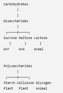
Carbohydrates

Monosaccharides
       |
Glucose, Fructose

Disaccharides
       |
Sucrose, Maltose, Lactose

Polysaccharides
       |
Starch, Cellulose, Glycogen
Important Facts
Glucose
- Aldohexose
- Formula: C₆H₁₂O₆
- Exists in α and β forms
- Forms pyranose ring
Fructose
- Ketohexose
- Fruit sugar
- Forms furanose ring
Important Disaccharides
| Compound | Components | Nature |
|---|---|---|
| Sucrose | Glucose + Fructose | Non-reducing |
| Maltose | Glucose + Glucose | Reducing |
| Lactose | Galactose + Glucose | Reducing |
Important Polysaccharides
| Polysaccharide | Function |
|---|---|
| Starch | Plant storage carbohydrate |
| Glycogen | Animal storage carbohydrate |
| Cellulose | Plant structural material |
2. Proteins
Basic Unit:
Amino acids
General structure:
      H
      |
H₂N — C — COOH
      |
      R
Peptide Bond
The bond between two amino acids:−CO−NH−
is called peptide linkage.
Protein Structure Levels
Primary Structure
Sequence of amino acids.
Secondary Structure
- α-helix
- β-pleated sheet
Tertiary Structure
Three-dimensional folding.
Quaternary Structure
Arrangement of multiple chains.
Protein Classification
Fibrous Proteins
- Long chain structure
- Insoluble
- Example: Keratin
Globular Proteins
- Spherical shape
- Usually soluble
- Example: Insulin
Denaturation
Loss of biological activity of protein due to:
- Temperature change
- pH change
Primary structure remains unchanged.
3. Enzymes
Definition
Enzymes are biological catalysts that increase the rate of biochemical reactions.
Properties
- Mostly proteins
- Highly specific
- Reduce activation energy
- Work under mild conditions
Enzyme Action
Enzyme + Substrate
        ↓
Enzyme-Substrate Complex
        ↓
Product + Enzyme
4. Vitamins
Classification
Vitamins

Fat Soluble:
A, D, E, K

Water Soluble:
B-group, C
Important Deficiency Diseases
| Vitamin | Deficiency |
|---|---|
| A | Night blindness |
| B₁ | Beriberi |
| B₁₂ | Pernicious anaemia |
| C | Scurvy |
| D | Rickets |
| K | Increased clotting time |
5. Nucleic Acids
Types
- DNA
- RNA
Nucleotide
Contains:
- Nitrogenous base
- Sugar
- Phosphate group
Nucleoside
Contains:
- Nitrogenous base
- Sugar
(No phosphate)
DNA
Function:
- Stores genetic information
- Transfers hereditary characters
Base Pairing:
A = T
G ≡ C
RNA
Types:
| RNA Type | Function |
|---|---|
| mRNA | Carries genetic message |
| tRNA | Transfers amino acids |
| rRNA | Forms ribosomes |
Important Differences for Exams
DNA vs RNA
| DNA | RNA |
|---|---|
| Deoxyribose sugar | Ribose sugar |
| Double stranded | Usually single stranded |
| Thymine present | Uracil present |
| Genetic storage | Protein synthesis |
Final Biomolecules Mind Map
                 BIOMOLECULES

                      |
 ------------------------------------------------
 |              |              |                |
Carbohydrates Proteins     Nucleic Acids    Vitamins
 |              |              |                |
Sugars       Amino acids      DNA/RNA        A,D,E,K
 |              |                               B,C
Energy       Peptide bonds
Storage      Protein structure